
Imagine being the first person to place a flea beneath a lens and discover that the irritating little speck was wearing articulated armor. Its claws curved like grappling hooks. Its body bristled with sensory hairs. Its familiar jump suddenly belonged to an animal of astonishing mechanical complexity. The flea had always been magnificent. Humanity had simply lacked the glass to see it.
In 1665, Robert Hooke pulled that curtain back for the public. His Micrographia, the first fully illustrated book devoted to microscopy, transformed a flea into a page-sized revelation and described microscopy as opening “a new visible World.” Eleven years later, Antoni van Leeuwenhoek reported living “animalcules” in water in a remarkable 1676 letter to Henry Oldenburg. In 1687, his observations of ant eggs and larvae challenged what people thought they already knew about ant development.
A microscope does more than enlarge. It reminds us that reality is not limited to what the unaided eye can perceive. Four centuries later, choosing the right instrument still begins with one deceptively simple question: Do you want to look at a whole insect, or look through a thin piece of one?
This countdown begins with a focused slide collection at #5 and works toward our favorite at #1. Along the way, we will separate stereo microscopes from compound microscopes, explain what magnification can and cannot do for identification, and pull the curtain back on five very different ways to look closer.
Quick answer
Our top pick is the portable 20X/50X binocular stereo microscope set. Its useful working distance, reflected LED lighting, two magnification options, and included specimens make it the most approachable choice here for examining whole insects and other opaque objects. The continuous-zoom trinocular microscope is the more specialized bench instrument, while the three prepared slide collections are best for readers who already own a compatible microscope with transmitted illumination.
How we chose
We compared the five supplied products by what they actually are, not by the wording of their marketplace listings. We considered intended use, optical design, magnification, working distance, lighting, specimen format, slide material, storage, camera capability, setup requirements, and the kind of insect detail each option could reasonably reveal.
We also ranked complete viewing instruments separately from specimen accessories. A box of prepared slides may be fascinating, but it is not a microscope, and the type of illumination available matters as much as the magnification printed on the package.
Product details, availability, and shipping restrictions can change. Confirm current contents, specifications, and requirements with the retailer before purchase.
Reviewed by Carol Maier, B.Sc., professional entomologist and founder of the original Victoria Bug Zoo.
Affiliate disclosure: As an Amazon Associate I earn from qualifying purchases. This article contains affiliate links. If you make a purchase through one of these links, Bug Zoo® may earn a commission at no additional cost to you.
Two microscopes, two kinds of seeing
People often use “microscope” as though it describes one instrument. It does not. A stereo microscope and a compound microscope solve different visual problems, and an entomologist may need both.
The stereo microscope: depth, working room, and whole insects
A stereo microscope, often called a dissecting microscope, sends a slightly different view to each eye through two separate optical pathways. The brain fuses those views into an image with real depth. Magnification is usually lower than it is in a compound microscope, but the field of view and working distance are larger. That leaves room beneath the objective for forceps, a pin, a brush, or an intact insect.
Whole beetles, moth wings, antennae, tarsi, mouthparts, setae, wing venation, and other opaque structures are commonly illuminated from above. Nikon notes that opaque specimens usually benefit from reflected light, while transparent specimens generally respond better to transmitted illumination.
True stereoscopic depth arrived much later than the earliest compound microscopes. By 1897, ZEISS recorded its Greenough stereomicroscope, a design based on paired, inclined optical paths. That same principle still gives modern dissecting microscopes their wonderfully useful three-dimensional view.
The compound microscope: transmitted light and finer internal detail
A compound microscope uses objective lenses to produce much higher magnification through a shared optical axis. Even when it has two eyepieces, it does not automatically produce a stereo view. In conventional brightfield work, light rises from beneath the stage and passes through a thin specimen before entering the objective. Transmitted brightfield illumination is well suited to thin, stained biological material such as tissue sections and mounted insect parts.
That is where prepared slides belong. A mounted mosquito wing, leg, scale, or tissue section can reveal fine structures under transmitted light, but a thick, opaque beetle is another matter. It blocks the light, offers little working room, and may sit outside the useful depth of focus. For the intact animal, reach for the stereo microscope. For a thin, transparent preparation, reach for the compound microscope.
Magnification is not the same as identification
A closer look can reveal diagnostic characters that the naked eye misses: the number and shape of antennal segments, arrangements of setae, tarsal claws, wing veins, scales, punctures, mouthparts, and tiny proportions between structures. But magnification does not name the insect for you.
Reliable identification still depends on the condition and orientation of the specimen, the correct taxonomic key, comparison material, geographic information, life stage, and sometimes dissection. The best microscope is not necessarily the one with the largest number printed on the box. It is the one that shows the character you actually need to see.
#5: Focused 10-Slide Whole-Insect Collection

This compact collection concentrates on 10 prepared insect specimens and life stages. The supplied list includes examples such as mosquito, aphid, ant, fruit fly, and housefly material, giving learners a focused alternative to slide boxes filled mostly with plants and mammalian tissues.
Whole mounts can be especially satisfying because the viewer can relate a familiar insect shape to its finer structures. They can also be optically challenging. Thickness, mounting quality, and overlapping body parts may keep every layer from coming into focus at once, so the most useful view will depend on the specimen and microscope.
Why it stands out
- The subject stays tightly focused on insects rather than treating them as a small part of a general biology assortment.
- The optical-glass slides come in a compact storage box measuring approximately 1 by 3 inches.
Good to know: This is a prepared slide set, not a microscope. It is normally most useful with a compound microscope that provides transmitted light, although compatibility also depends on specimen thickness.
Best for: Students who already have a compatible microscope and want a small, insect-centered collection.
#4: 30-Slide Glass Biology Collection

This broader collection includes more than 30 prepared glass slides featuring plants, algae, mammalian tissues, cells, and insect parts. Its value lies in comparison. An insect structure becomes more meaningful when a learner can place it within a larger survey of biological forms.
The slides use the standard 25 by 75 millimeter format and are described as having polished, rounded edges. Because these are thin biological preparations, a compatible compound microscope with transmitted light is the natural partner.
Why it stands out
- The collection places insect material within a wider introduction to cells, tissues, plants, algae, and animals.
- Standard-size glass slides should suit many conventional compound microscope stages.
Good to know: The supplied information describes prepared slides only. A microscope is not included.
Best for: Homeschool study, beginners, and teaching demonstrations that compare insect anatomy with wider biology.
#3: Continuous-Zoom Trinocular Stereo Microscope

This specialized bench microscope has a stated 0.7X to 5X zoom objective and 7X to 50X total magnification. Continuous zoom lets the observer move from a wider view of the whole specimen to a closer examination of one structure without repeatedly swapping eyepieces.
The trinocular head adds a third optical port intended for a camera, while WF10X23 eyepieces provide the listed viewing field. An LED ring light illuminates opaque subjects from above. The supplied accessories lean heavily toward electronics repair, including a soldering exhaust fan, work floor, and silicone trays, but optics do not care whether the object below them is a circuit board or a beetle's tarsus.
Why it stands out
- Continuous zoom supports smooth movement across the stated 7X to 50X magnification range.
- The trinocular format creates a route to photography or video when paired with compatible camera equipment.
Good to know: The working distance varies with the microscope version and the position of any Barlow lens. The supplied configuration is designed mainly as a repair workstation, and a camera is not identified as included.
Best for: Experienced hobbyists who want zoom observation, reflected ring lighting, and a camera-ready bench setup.
#2: 48-Slide Introductory Biology Collection
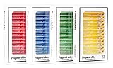
With 48 prepared slides covering animals, insects, plants, flowers, and tissue samples, this is the broadest specimen collection in the countdown. Labeled preparations make it easier for a new learner to connect what appears in the eyepiece with a named biological source.
The supplied record describes high-transparency plastic slides with smooth edges and storage intended to protect them from moisture. The format favors breadth over an exclusively entomological focus, making it useful for a young naturalist whose questions refuse to stay inside one phylum.
Why it stands out
- Forty-eight labeled preparations create the widest introductory survey in this group.
- The collection includes insect parts alongside plant, animal, flower, and tissue material.
Good to know: This product is a prepared slide collection and requires a compatible microscope, normally a compound model with transmitted light.
Best for: Young learners, family educators, and classrooms building a broad introductory biology library.
Now pull the curtain all the way back. Our top pick is the option that lets a new observer place a whole insect beneath the lens and begin exploring.
#1: Portable 20X/50X Binocular Stereo Microscope Set

This portable stereo microscope offers 20X and 50X viewing through WF10X and WF25X eyepieces paired with a 2X objective. Its adjustable working range of 65 to 102 millimeters leaves useful space for larger, opaque specimens, while binocular viewing and adjustable interpupillary distance accommodate different users.
Reflected LED lighting brightens the specimen from above, and the portable design runs on two AA batteries. The supplied listing also includes insect specimens, 10 slides, and 12 mineral specimens, so a beginner has several objects ready for a first look.
Why it takes our top spot
- The 65 to 102 millimeter working range supports whole-insect observation and simple manipulation beneath the objective.
- Two eyepiece choices provide stated 20X and 50X magnification without a complex zoom system.
- Included insect and mineral specimens make the first session easier to begin.
Good to know: The LED light requires two AA batteries, which are not included. The listing also mentions prepared slides, but do not assume every slide will perform well without illumination from beneath.
Best for: Families, classrooms, and beginners who want an approachable stereo microscope for studying whole insects and other natural objects.
Final thoughts
The portable 20X/50X binocular stereo microscope is our best overall pick for someone who wants to look closely at whole insects without building a laboratory first. Choose the continuous-zoom trinocular model for a more specialized camera-ready bench instrument, the 48-slide collection for the broadest introduction to biology, the 30-slide glass set for conventional classroom preparations, or the focused 10-slide collection when insects are the only stars you want on stage.
Hooke's flea did not become extraordinary when he magnified it. The lens merely forced human eyes to catch up with the reality that had been there all along. That may be the most beautiful thing a microscope teaches: wonder is not manufactured. Sometimes it is waiting just beyond the limits of our sight.
Frequently asked questions
What is the difference between a stereo microscope and a compound microscope?
A stereo microscope uses two optical paths to provide depth, lower magnification, a larger field of view, and more working room. It is especially useful for whole, opaque insects illuminated from above. A compound microscope provides higher magnification through a shared optical axis and commonly sends light through a thin specimen mounted on a slide.
Can a stereo microscope view prepared slides?
Some stereo microscopes can view transparent specimens when equipped with transmitted illumination, but many basic models illuminate only from above. Thin biological slides are normally better suited to a compound microscope with light passing through the preparation.
What magnification is useful for identifying insects?
For many whole-insect characters, a broad low-power range around 10X to 40X is more useful than extreme magnification because it preserves depth, field of view, and working distance. Smaller structures may require more magnification, but the correct taxonomic character determines the instrument and setting.
What does trinocular mean on a microscope?
A trinocular microscope has two eyepieces for observation plus a third optical port intended for a compatible camera. The third port can support photography, video, teaching displays, and documentation, but the camera and adapter may be sold separately.
Can a microscope identify an insect automatically?
No. A microscope reveals characters that may be needed for identification, but a reliable name still depends on a suitable key, specimen condition, life stage, locality, comparison material, and sometimes expert examination or dissection.
Thanks for supporting our mission to make the world a better place for everyone, bugs included.